
About the project


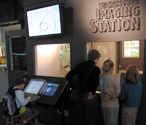
visitors using Microscope Imaging Station
In Summer 2004, the Exploratorium launched the most ambitious microscope facility ever created for use by the general public, the Microscope Imaging Station. The initial phase of the project gives visitors the ability to image living specimens, as well as control the microscopes themselves.
At the museum, you can select among various specimens, move over them, change the magnification and focus, and, where appropriate, change the lighting to illuminate the specimen or use reflected light and fluorescence to dramatically change how the specimens look.
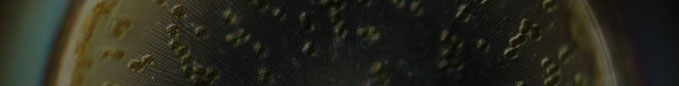
The images you see here have been created at the Microscope Imaging Station. They reflect the diversity of topics and specimens you might see on a typical visit.
A primary goal of our facility is to open a door to the wonder of the microscopic world and allow you to explore it. By empowering you with the instruments to explore this unfamiliar universe, we seek to recreate some of the excitement and wonder that the earliest biological researchers found as they discovered another world all around them.
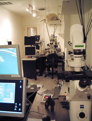
inside the Microscope Imaging Station
The development of the Microscope Imaging Station began in 2000 with funding from the National Center for Research Resources (a center within the National Institutes of Health) and the David and Lucile Packard Foundation.
An array of research scientists joined us in developing the Microscope Imaging Station, most notably Christian Sardet. A French cell biologist with a passion for imagery and a love of teaching, Dr. Sardet helped in the conception and development of the project.
We developed the software and wrote our own programs to control stage movement (for specimen positioning), focus, specimen selection, magnification, and lighting. We also developed interactive multimedia to guide visitors through the exploration of samples.
Numerous biomedical researchers and their laboratories across the United States have helped with intellectual input and specimens.
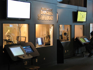
south end of
Microscope Imaging Station
In addition to using some of the latest devices in microscope technologies, the Imaging Station also provides a window on revolutionary research techniques. For example, in 1994, Columbia University's Martin Chalfie inserted a gene for a fluorescent jellyfish protein into a bacterium (E. coli) and a roundworm (C. elegans) and found that the genetically-modified organisms emitted an eerie green glow under certain lighting conditions.
Other scientists built on this technique to create a powerful tool that makes hidden structures and processes easier to study.
The coupling of vital staining with this green fluorescent protein (GFP) gene allows scientists to observe events inside developing cells and detect the presence of diseased structures and environmental toxins with extreme sensitivity. This newly created sensitivity has sparked new insights and discoveries, re-revolutionizing the capabilities of the light microscope.
At the Imaging Station, you can take advantage of this technique to observe specific types of cells such as the brain cells, sex cells, or muscle cells of roundworms. You can also peer inside the developing embryo of a tropical zebrafish whose circulatory cells have been made visible by the protein made from the transplanted GFP gene.
Over the next year, we intend to add a major component to the Microscope Imaging Station on mouse stem cells and the process of differentiation.
We hope you have the opportunity to use this unique facility and its images to better understand living things, human health, and disease.