
Classroom Explorations:
What's the Size of What You See?
Sea Urchin Embryo Cell Division
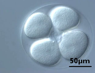
Use the scale bar in this image as a reference when you watch the video below.
The video, Sea urchin embryo cell division, shows the first 90 minutes of sea urchin (
Lytechinus pictus
) embryonic development. Nuclei are visible as “thumbprints“ in each cell. Microtubules are faintly visible just prior to and during mitosis. (The elapsed time is about two hours.)

Sea urchin eggs were fertilized, then immediately mounted in seawater between a slide and coverslip (using a silicon spacer). Images were taken at room temperature on a compound inverted microscope using a 40x DIC objective and digital camera.