
Neurons: A fish-eye view of the brain
Navigating a maze of cells
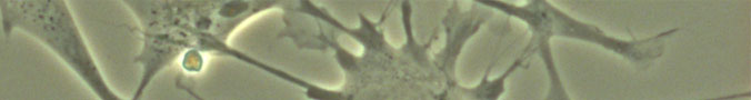
The vast number of neuronal connections inside a brain creates a network of almost unfathomable complexity. Brain function relies on this intricate maze; it’s what creates the processing power that allows a brain to accomplish complicated tasks like responding to a visual cue, moving a body part, or (in humans) recognizing a familiar face and saying “hello.”
But how exactly does the brain get wired up? The way this impressive network grows and organizes itself during development (and, sometimes, during adulthood) is a puzzle many researchers are trying to solve.
So far, scientists know that the number of neurons in an individual’s brain is determined by genetics—but how those neurons are wired together, and the number of synapses among them, is influenced by environment. By making movies of zebrafish neurons as they develop, scientists in the laboratory of Stanford neurobiologist Stephen Smith are getting glimpses of how this process unfolds.
Learning from a fish
Why study fish brains? Zebrafish have several qualities that make them good models for studying neuron development. They’re small, they're inexpensive, and they grow and reproduce quickly. They also have enough interesting behaviors to make them useful to observe, but not so many that they are confusing.
Another advantage is that their brains develop rapidly: A zebrafish grows from single cell to swimming larvae with a functional brain in under three days. In humans, this same process takes months. This compressed time scale allows scientists to track the entire wiring process of the fish's brain. An extra bonus is that zebrafish are transparent to certain wavelengths of light, so researchers can easily see what’s happening inside them as they grow.
Scientists are also able to engineer zebrafish with specific genetic characteristics. In Smith's lab, for example, they’ve modified the fish’s genome to actually illuminate particular neurons in a living fish’s brain. The fish’s transparency allows the researchers to bombard it with a powerful laser (affectionately known as the “death ray”). This light would burn opaque organisms, but shines right through the zebrafish. Using these techniques, scientists can watch and measure the growth, development, and activity of specific neurons without harming the living fish.


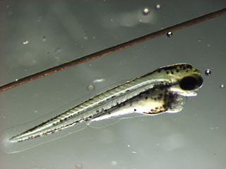
A three-day-old zebrafish next to a human hair.
Researchers in the Smith lab study neurons developing in the optic system of newly-hatched zebrafish. When a fish is three days old, it begins to see. Researchers put it in a warm bath, under a microscope, and show the tiny fish patterns on a miniature LCD screen. They've found that the fish's brain is most active when the screen's moving spots are the same size as its favorite food. These results imply that neurons in the zebrafish’s visual system are programmed to detect certain movements more readily than others, which may help the young fish find food early in life.
Next: Speed-dating neurons make connections »